Services

1. Routine Tissue Examination and Diagnosis
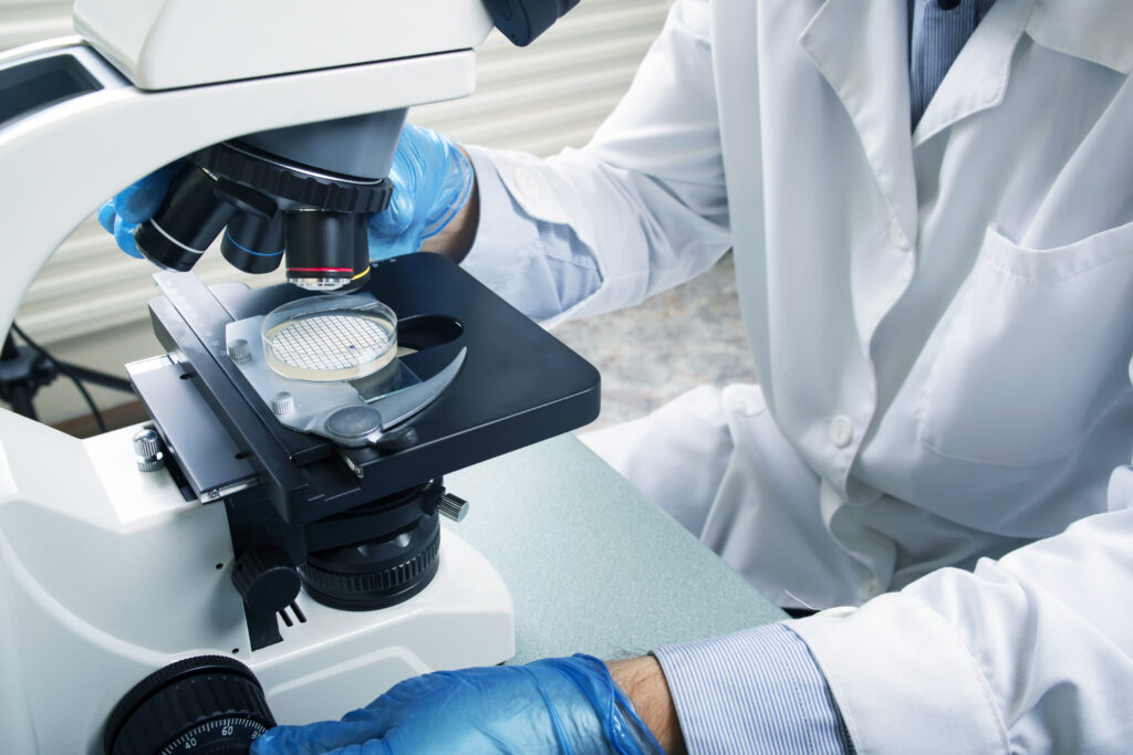
This service constitutes the majority of our work and generally includes gross and microscopic examination and diagnosis of oral, head, and neck surgical biopsy and excision specimens. (See separate Obtaining Maximally Diagnostic Oral Tissue Specimens). Rarely, certain specimens are only examined grossly—primarily for medicolegal identification purposes. Special histochemical stains are performed when needed to further identify cell products, exogenous materials, or microorganisms. Immunohistochemical stains are utilized when needed to identify the histogenesis or to classify poorly differentiated neoplasms. These special studies take longer than the usual overnight turnaround time for uncomplicated specimens, and additional charges are incurred.
2. Direct Tissue Immunofluorescence Examination
This highly specific analysis is used primarily for the diagnostic confirmation of immunologically mediated vesiculobullous diseases, such as mucous membrane (cicatricial) pemphigoid and pemphigus vulgaris. However, it can also be helpful in the confirmation of atypical cases of oral lichen planus as well as lupus erythematosus and erythema multiforme. Because some oral conditions (i.e. dysplasia, squamous cell carcinoma, etc.) cannot be evaluated with fluorescence microscope, immunofluorescence specimens must be accompanied by a routine specimen (submitted in formalin for hematoxylin and eosin staining), if a recently obtained specimen of the same disease process is not available for concurrent microscopic examination. Specimens should be submitted in unexpired immunologic transport medium (Michel’s solution or Zeus fixative). The specimen will forwarded to a referral laboratory that will process and diagnose the immunofluorescent studies. The patient will receive a separate bill from our referral laboratory. Because of the expense and limited shelf life of the transport medium, it should be ordered from the lab by telephone approximately 2 weeks before a patient’s biopsy appointment.
3. Microslide Review/Second Opinion
This service is being used with increasing frequency because of the increased utilization of managed healthcare plans. The high volume of cases processed by the contracted pathology labs and the usual lack of an oral pathologist on their staff can result in more general/nonspecific diagnoses of oral, head, and neck specimens than clinicians and patients desire. However, our expertise with the oral cavity and Head & Neck regions usually allows us to provide much more specific histopathologic diagnoses, which facilitates more precise care.